Antimicrobial Peptides and Interleukins in Cleft Soft Palate
DESCRIPTION
Cleft palate is one of the most common and well-studied congenital anomalies; however, the role of protective tissue factors in its pathophysiology is still debated. The aim of our study was to evaluate interleukin and antimicrobial peptide appearance and distribution in cleft palate. Eight soft palate samples were obtained during veloplasty procedures.
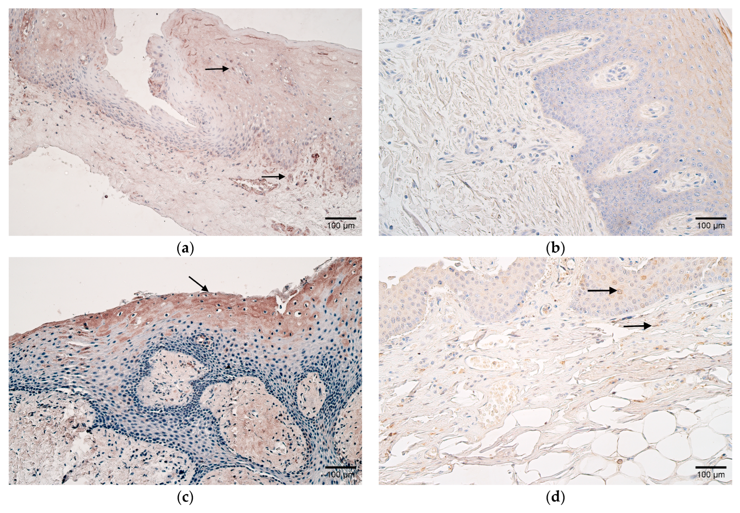
Immunohistochemical staining was applied to detect HBD-2-, HBD-3-, HBD-4-, LL-37-, IL-10-, and CD-163-positive cells via light microscopy. For statistical evaluation, the Mann–Whitney U test and Spearman’s rank correlation coefficient were used.
A significant difference between study groups was observed for HBD-2 and IL-10 in epithelial and connective tissue as well as HBD-4 in connective tissue. The number of HBD-3-positive cells was moderate in the patients, and few were observed in the controls. The number of LL-37-positive cells varied from a moderate amount to a numerous amount in both study groups, whilst CD-163 marked a moderate number of positive cells in patients, and a few-to-moderate amount was observed in the controls.
Numerous correlations between studied factors were revealed in cleft tissues. The increase in antimicrobial peptides HBD-2 and HBD-4 and anti-inflammatory cytokine IL-10 suggested a wide compensatory elevation of the local immune system against cleft-raised tissue changes. The correlations between the studied factors (HBD-2, HBD-3, HBD-4, LL-37, and IL-10) proved the synergistic involvement of common local defense factors in postnatal cleft palate morphopathogenesis.