Characterization of Macrophages and TNF-α in Cleft Affected Lip Tissue
DESCRIPTION
Orofacial clefts are one of the most common congenital anomalies worldwide; however, morphopathogenesis of the clefts is not yet completely understood.
Taking the importance of innate immunity into account, the aim of this work was to examine the appearance and distribution of macrophages (M) 1, M2, and TNF-α, as well as to deduce any possible intercorrelations between the three factors in cleft affected lip tissue samples.
Twenty samples of soft tissue were collected from children during plastic surgery. Fourteen control tissue samples were obtained during labial frenectomy.
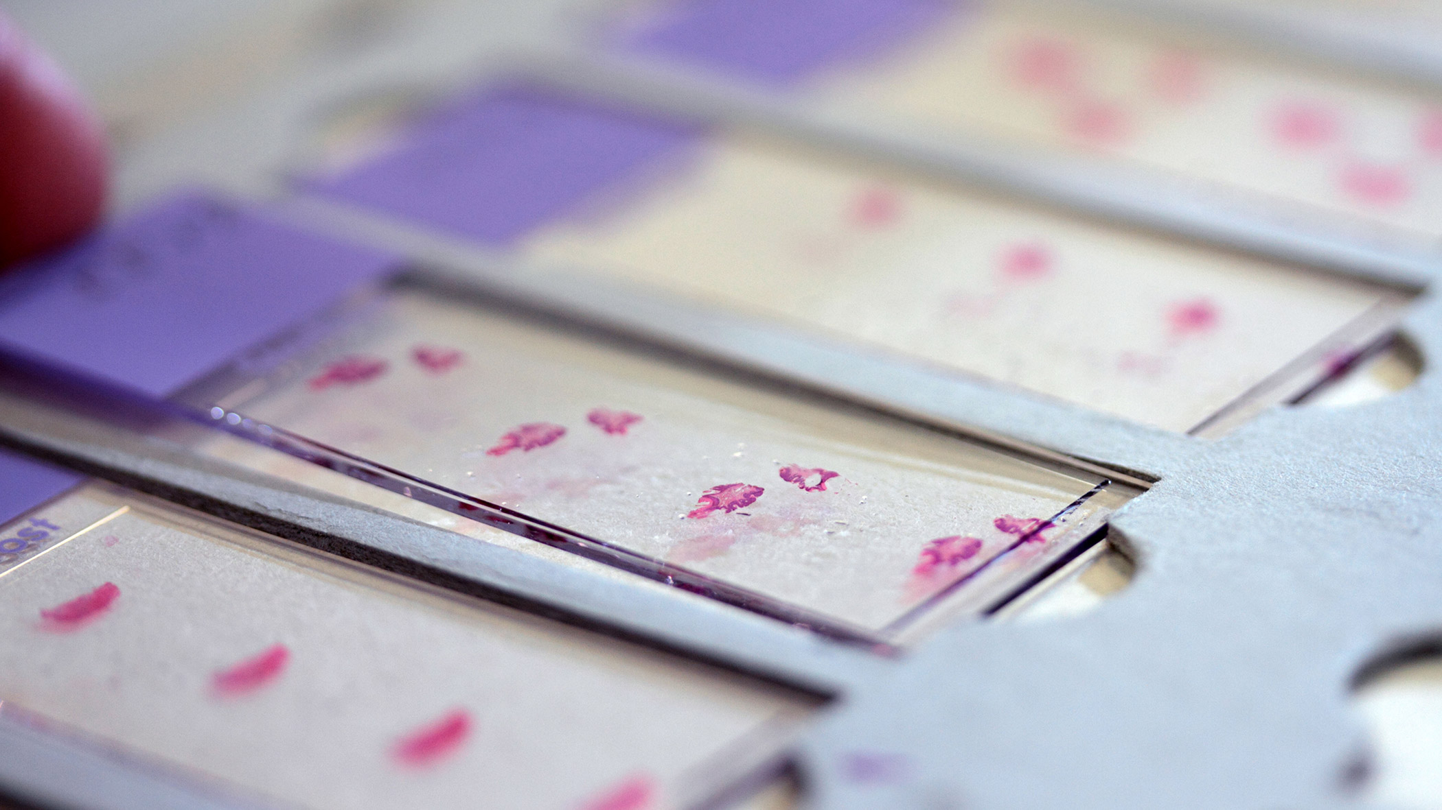
Tissues were immunohistochemically stained, analysed by light microscopy using a semi-quantitative method, and the Mann–Whitney U and Spearman’s tests were used to evaluate statistical differences and correlations. A statistically significant difference in the distribution was observed only in regard to M1.
A weak correlation was observed between M2 and TNF-α but a moderate one between M1 and M2 as well as M1 and TNF-α.
However, only the correlation between M1 and M2 was statistically important.
The rise in M1, alongside the positive correlation between M1 and TNF-α, suggested a more pro-inflammatory/inflammatory environment in the cleft affected lip tissue.
The moderate positive correlation between M1 and M2 indicated an intensification of the protective mechanisms.