The Mechanistic Study of Mitochondrial Autophagy and Ferroptosis in the Progression of Decreased Ovarian Reserve
DESCRIPTION
Abstract
Objective
To investigate the mechanism in the progression of decreased ovarian reserve (DOR).
Methods
Three-month-old female SD rats were employed and randomly divided into the model group and the normal group. The model group was intraperitoneally injected with 4-vinylcyclohexene diepoxide (VCD). Thereafter, blood sample from the abdominal aorta was taken, and rats were sacrificed, and ovarian tissues were obtained by laparotomy.
Results
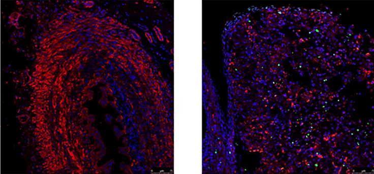
HE staining results revealed that the model group exhibited significantly reduced ovarian volume, increased follicular atresia, and decreased quantities of growing follicles and corpus luteum, thereby indicating degraded reserve function of ovarian. TEM images revealed that prominent autophagic vacuoles could be observed in the model group, accompanied by the mitochondria shrinkage and generation of the autophagosome. The expression of Pink1, Parkin, BNIP3L and LC3II genes in ovaries of the model group was significantly higher than those of the normal group (P < 0.05). In addition, the protein expression of Pink1, Parkin, BNIP3L and LC3II in ovaries of the model group were higher than those of the normal group (P < 0.05). The expression of Fe2+ and GSH in oocytes of the model group was higher than those of the normal group (P < 0.05). The expression of FTH1 and GPX4 in oocytes of the model group was significantly higher than those of the normal group (P < 0.05).
Conclusions
Mitochondrial autophagy and ferroptosis may participate in the progression of decreased ovarian reserve (DOR).