The Neuronal nitric oxyde synthase positive neurons
DESCRIPTION
The study was designed to analyze the Neuronal nitric oxyde synthase positive neurons present in the indusium griseum by describing their distribution and morphology.
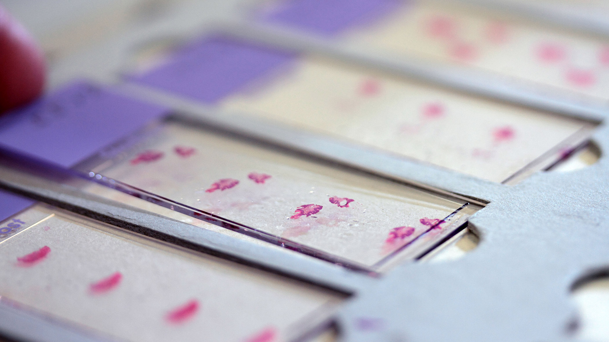
To this purpose, sagittal serial sections from paraffin or frozen autopsy specimens of corpus callosum were processed by immunohistochemistry and immunofluorescence, using an antibody against the neuronal form of the enzyme nitric oxyde synthase.
To test the specificity of the antibody used, Western Blot was performed in the indusium griseum of the same specimens.
The stainings revealed the presence of many neuronal nitric oxyde synthase-immunopositive neurons in human indusium griseum, located along both rostral-caudal and medio-lateral directions.
In particular, they were more numerous 1 mm apart from the midline, and their number peaked over the body of the corpus callosum.
They showed different morphologies; in some cases, they were located at the boundary between indusium griseum and corpus callosum.
More densely packed in proximity to the pial arteries penetrating into the corpus callosum.
The significant presence and distribution of neuronal nitric oxyde synthase-immunopositive neurons suggests that indusium griseum likely plays a functional role in the neurovascular regulation within the corpus callosum.
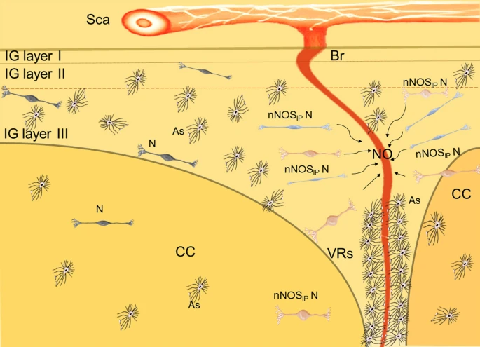
The Neuronal nitric oxyde synthase positive neurons surround the arterioles and control the vasomotore tone secreting nitric oxyde.
Two morphological types of nNOSIP N can be appreciated by the use of different colors: fusiform (blue) and ovoidal (pink).
Also NeuN-immunopositive neurons and many astrocytes are present, more numerous in IG than in CC.
Here the Schematic representation of human adult IG and the neurovascular unit originating from sopracallosal artery (Sca) that branches into smaller arterioles: Picture.
